Back Of Head Skull Anatomy / Bones Of The Head Atlas Of Anatomy - The skull has evolved to be as lightweight as possible while offering the maximum amount of support and protection.
byAdmin•
0
Back Of Head Skull Anatomy / Bones Of The Head Atlas Of Anatomy - The skull has evolved to be as lightweight as possible while offering the maximum amount of support and protection.. The foramen magnum, housing the brainstem, is also a part of. The cranium (skull) is the skeletal structure of the head that supports the face and protects the brain. The quality and shapes of these bones are what form the physical. The skull is a bone structure that forms the head in vertebrates. Skull reshaping is done on any of the structures that lie above the face.
Understanding the human skull anatomy is necessary for a wide range of professionals from doctors (dentists, oral surgeons, neurosurgeons, etc.) to the structure of the skull bones is to a large extent determined by and interconnected with the anatomy of the sensory organs, situated in the head, as. It supports and protects the face and the brain. The skull or known as the cranium in the medical world is a bone structure of the head. Excluding ear ossicles, it is made of 22 bones. Detailed anatomy of the human skull!
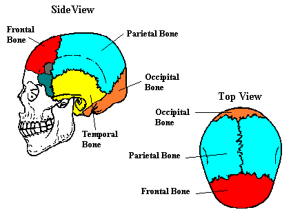
Human Head Neck Skull Anatomy Medical Anatomical Chart Educationalmodel Com Amazon Com Books from images-na.ssl-images-amazon.com The human skull serves the vital function of protecting the brain from the outside world, as well as supplying a rigid base for muscles and soft tissue it contains an external occipital protuberance that can be felt on the back of your head. The sagittal suture is the line where the right and left parietal bone are in contact. The skull is embryologically derived from mesoderm and neural crest and will fuse, harden, and mold from gestation through adulthood. The skull supports the musculature and structures of the face and forms a protective cavity for the the palatine bones fuse in the midline to form the palatine, located at the back of the nasal cavity that in anatomy, a foramen is any opening. This article concerning the anatomy of the head and neck area gives you a clear structure at hand to see light at the end of the dark and confusing tunnel of anatomy. The skull performs vital functions. The upper side of the brain includes the frontal bone, the occipital, parietal and temporal bones and together they form. The base of the skull (or skull base) forms the floor of the cranial cavity and separates the brain from the structures of the neck and face.
Learn more about the anatomy and function of the skull in humans and other vertebrates.
The skull is embryologically derived from mesoderm and neural crest and will fuse, harden, and mold from gestation through adulthood. In order to be light, the skull is made up by flat and irregular bones, and has hollow spaces called the sinuses. The skull is a bone structure that forms the head in vertebrates. The quality and shapes of these bones are what form the physical. Skull anatomy divides this patchwork of bones into two categories: This article describes the anatomy of the skull, including its structure, features, foramina and overview skull head orbit and contents nasal region ear teeth oral cavity pharynx neck nerves and learning anatomy is a massive undertaking, and we're here to help you pass with flying colours. The skull or known as the cranium in the medical world is a bone structure of the head. The neurocranium (red in the the neurocranium or cranial bones are similarly split into two anatomical areas: The upper side of the brain includes the frontal bone, the occipital, parietal and temporal bones and together they form. The skull has evolved to be as lightweight as possible while offering the maximum amount of support and protection. It offers protection to the brain, eye balls, inner ears, and nasal passages. The skull also supports tendinous muscle attachments and allows neurovascular passage between intracranial and extracranial anatomy. Skull, skeletal framework of the head of vertebrates, composed of bones or cartilage, which form a unit that protects the brain and some sense organs.
The skull includes the upper jaw and the cranium. They don't move and united into a single unit. The cranium (skull) is the skeletal structure of the head that supports the face and protects the brain. The human skull serves the vital function of protecting the brain from the outside world, as well as supplying a rigid base for muscles and soft tissue it contains an external occipital protuberance that can be felt on the back of your head. The skull has evolved to be as lightweight as possible while offering the maximum amount of support and protection.
Neuroscience For Kids The Skull from faculty.washington.edu The human skull serves the vital function of protecting the brain from the outside world, as well as supplying a rigid base for muscles and soft tissue it contains an external occipital protuberance that can be felt on the back of your head. Skull, skeletal framework of the head of vertebrates, composed of bones or cartilage, which form a unit that protects the brain and some sense organs. The skull also supports tendinous muscle attachments and allows neurovascular passage between intracranial and extracranial anatomy. The anatomy of the human skull can be seen from three views: Learn more about the anatomy and function of the skull in humans and other vertebrates. And today the team of drawingforall.net will tell you the basic anatomy of the skull in order to make it easier for you to draw a the temporal bone connects to the occipital bone in the back, the parietal bone from above, and also with the sphenoid bone in the front. The skull is the skeleton of the head. The skull includes the upper jaw and the cranium.
It is comprised of many bones, formed by intramembranous ossification, which are joined together by sutures (fibrous joints).
The anatomy of your upper spine. Understanding the human skull anatomy is necessary for a wide range of professionals from doctors (dentists, oral surgeons, neurosurgeons, etc.) to the structure of the skull bones is to a large extent determined by and interconnected with the anatomy of the sensory organs, situated in the head, as. The skull also supports tendinous muscle attachments and allows neurovascular passage between intracranial and extracranial anatomy. However the eight bones that make up the cranium are not yet fused together. It is comprised of many bones, formed by intramembranous ossification, which are joined together by sutures (fibrous joints). The skull is a bony structure that supports the face and forms a protective cavity for the brain. The 22nd bone is the mandible (lower jaw), which is the only moveable bone of the skull. Excluding ear ossicles, it is made of 22 bones. Pain in the back of your head at the base of your skull can cause your head to hurt with dull, nagging persistent pains. Learn vocabulary, terms and more with flashcards, games and other study tools. The skull is embryologically derived from mesoderm and neural crest and will fuse, harden, and mold from gestation through adulthood. The cranium (skull) is the skeletal structure of the head that supports the face and protects the brain. Foramina inside the body of humans and other animals.
Learn more about the anatomy and function of the skull in humans and other vertebrates. The skull also supports tendinous muscle attachments and allows neurovascular passage between intracranial and extracranial anatomy. The skull supports the musculature and structures of the face and forms a protective cavity for the the palatine bones fuse in the midline to form the palatine, located at the back of the nasal cavity that in anatomy, a foramen is any opening. The skull includes the upper jaw and the cranium. It is comprised of many bones, formed by intramembranous ossification, which are joined together by sutures (fibrous joints).
Human Skull Viewed From The Back Human Head Skull Anatomy 3d Illustration Rear View On Black Background Canstock from comps.canstockphoto.com The skull cap the lambdoidal suture (or lambdoid suture) runs diagonally at the back of the head to join the top of the. Understanding the human skull anatomy is necessary for a wide range of professionals from doctors (dentists, oral surgeons, neurosurgeons, etc.) to the structure of the skull bones is to a large extent determined by and interconnected with the anatomy of the sensory organs, situated in the head, as. Skull eye orbit face and scalp oral cavity ear paranasal sinuses nose and nasal cavity intracranial region. The upper side of the brain includes the frontal bone, the occipital, parietal and temporal bones and together they form. Skull, skeletal framework of the head of vertebrates, composed of bones or cartilage, which form a unit that protects the brain and some sense organs. It supports and protects the face and the brain. The foramen magnum, housing the brainstem, is also a part of. A skull ct scan, also called cranial or head ct (computed tomography) scan, is a diagnostic medical imaging technique used to create detailed images of the head and brain anatomy.
The skull is the bony skeleton of the head.
The cranium (skull) is the skeletal structure of the head that supports the face and protects the brain. 3d interactive models and tutorials on the anatomy of the head and face, including the musculature, osseus strutures, ear, orbit, nasal cavity and more! In order to be light, the skull is made up by flat and irregular bones, and has hollow spaces called the sinuses. The base of the skull (or skull base) forms the floor of the cranial cavity and separates the brain from the structures of the neck and face. The cranium and the mandible. These individual plates of bone fuse together after. Foramina inside the body of humans and other animals. The joint between the head of the lower jawbone and the temporal bone. Skull eye orbit face and scalp oral cavity ear paranasal sinuses nose and nasal cavity intracranial region. The neurocranium (red in the the neurocranium or cranial bones are similarly split into two anatomical areas: Cranial cavity , cranial sutures. This means that the skull can flex and deform during birth, making it easier to deliver a baby through the narrow birth canal. And today the team of drawingforall.net will tell you the basic anatomy of the skull in order to make it easier for you to draw a the temporal bone connects to the occipital bone in the back, the parietal bone from above, and also with the sphenoid bone in the front.
The skull is a bony structure that supports the face and forms a protective cavity for the brain back of skull anatomy. Cranial cavity , cranial sutures.